
PXE Guide: Eye
What Is PXE?
Pseudoxanthoma elasticum (PXE) is an inherited condition that causes abnormal mineralization in some elastic tissues of the body. Mineralization means that tiny deposits of calcium and other minerals build up where they do not usually belong.
PXE most often affects the skin, eyes, and mid-sized arteries. Less commonly, it may affect the gastrointestinal system. PXE was first recognized more than 100 years ago and much has been learned about it in recent decades.
Why Eye Care Matters in PXE
The eyes are among the main organs affected by pseudoxanthoma elasticum (PXE). In PXE, changes occur in the retina, the light-sensitive layer at the back of the eye.
Many PXE eye findings do not cause symptoms at first. Early changes are usually seen only during a dilated eye examination by an ophthalmologist or retina specialist.
PXE most often affects central vision, the vision used for reading, recognizing faces, driving, and seeing fine detail. Peripheral, or side, vision is usually preserved.
The Retina and Bruch’s Membrane
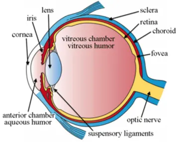
The eye works somewhat like a camera. Light enters the eye and is focused onto the retina. The retina sends visual information to the brain.
The macula is the central part of the retina. It is responsible for sharp central vision.
Behind the retina is a thin layer called Bruch’s membrane. Bruch’s membrane contains elastic tissue. In PXE, this layer can become mineralized and more fragile over time. These changes can lead to several eye findings seen in PXE.
Early Eye Findings
Peau d’orange
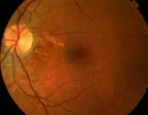 Peau d’orange means “orange peel” in French. In PXE, a mottled, orange-peel-like appearance is described in the retina. It is often one of the earliest eye signs of PXE and may appear in childhood or adolescence. It refers only to the eyes in PXE, not to skin.
Peau d’orange does not usually affect vision on its own. It is an important sign because it can help confirm a diagnosis of PXE.
Angioid streaks
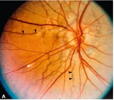 Angioid streaks are small breaks in Bruch’s membrane, the thin elastic layer behind the retina. During an eye exam, they may look like reddish, brownish, or gray lines spreading out from the area around the optic nerve.
Angioid streaks are sometimes compared to fine cracks in old lacquer or dried mud. The streaks themselves usually do not affect vision, but they can create places where fragile new blood vessels may grow, leak, or bleed.
Angioid streaks do not cause vision loss on their own. However, they matter because small abnormal blood vessels can sometimes grow through these breaks. These fragile new vessels may leak or bleed.
Comet lesions and drusen
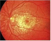 Some people with PXE have other retinal findings, including comet lesions or drusen. Comet lesions are yellowish or white retinal findings that can be helpful in diagnosing PXE. Drusen are small deposits under the retina and can occur in people with and without PXE.
A retina specialist can interpret these findings in the context of the full eye examination.
Choroidal Neovascularization
The most important PXE-related eye complication is choroidal neovascularization, often called CNV.
CNV means that small new blood vessels grow from the choroid, a blood-vessel-rich layer beneath the retina, through breaks in Bruch’s membrane. These new vessels are fragile and can leak or bleed.
When this happens near the macula, it can cause: • Wavy or distorted lines • Blurred central vision • A dark or missing spot in central vision • Difficulty reading • Difficulty recognizing faces • Sudden change in vision
CNV should be treated promptly. Early treatment gives the best chance of preserving vision. Check the Amsler Grid regularly for any distortion that might indicate CNV.
Treatment for Retinal Leaking or Bleeding The main treatment for PXE-related CNV is anti-VEGF injection therapy. These medicines are injected into the eye by a retina specialist to reduce leakage and bleeding from abnormal blood vessels. Several anti-VEGF medicines are used “off-label” by retina specialists, including aflibercept, ranibizumab, bevacizumab, faricimab, brolucizumab, and related biosimilars. Your retina specialist will decide which medicine and schedule are appropriate. They might call the medication by its trade name, such as Eylea® and Eylea HD® (aflibercept), Lucentis® and related ranibizumab biosimilars, Avastin® (bevacizumab, often used off-label in the eye), Vabysmo® (faricimab), and Beovu® (brolucizumab). Anti-VEGE options continue to evolve, largely through research and development in common retinal diseases such as AMD.
Anti-VEGF treatments have greatly changed the outlook for PXE eye disease. Many people with PXE can preserve useful vision when CNV is detected and treated early. These medicines were developed for AMD, so it is important that your ophthalmologist code them for macular degeneration so that insurance will pay for them. They are not approved, and never will be, for PXE because the market for these drugs is too small to justify the costs of regulatory (FDA, EMA, and so on) approval.
New retinal treatments are being developed rapidly, especially for age-related macular degeneration. These include longer-acting anti-VEGF drugs, higher-dose anti-VEGF drugs, medicines that target more than one pathway, refillable drug-delivery systems, complement inhibitors for geographic atrophy, and early gene-therapy approaches intended to reduce the need for frequent injections.
These treatments are being developed mainly for AMD, not PXE. Some may become relevant to PXE-related retinal bleeding because both conditions can involve abnormal leaking blood vessels under the retina. However, PXE is a different condition, and treatment decisions should be made by a retina specialist based on the individual eye findings.
Older treatments, such as laser photocoagulation, photodynamic therapy, and retinal transplant, were used in the past but are no longer a treatment for PXE-related CNV. If your ophthalmologist recommends one of these outdated treatments, get a second opinion or contact PXE International.
The Amsler Grid
An Amsler grid is a simple checkerboard-like tool used to check central vision. It can help a person notice changes that may suggest retinal leaking or bleeding. People with PXE should ask their retina specialist how often to use an Amsler grid and how to use it correctly. Or you can download one from the PXE International site here.
The Amsler grid does not replace regular eye exams. It is an early warning tool. Download one here.
Regular Eye Care
People with PXE should have regular dilated eye examinations, usually once a year. Many people with PXE are followed by a retina specialist, especially once angioid streaks are present.
Your eye doctor may use: • Dilated retinal examination • Retinal photography • Optical coherence tomography, or OCT • Fluorescein angiography, if leaking or bleeding is suspected • Other retinal imaging as needed
Fluorescein angiography and other specialized tests are not necessarily needed at every visit. They are usually used when the retina specialist needs more information.
When to Contact an Eye Doctor Urgently
Contact your retina specialist or eye doctor promptly if you notice: • Sudden distortion in central vision • Lines that should be straight (graph paper, bathroom tile) looking wavy or bent • A new dark, gray, blurry, or missing spot • Sudden trouble reading • Sudden change in vision in one eye
Do not wait for the next scheduled appointment. PXE-related retinal bleeding can often be treated, but timing matters.
Red Eye, Flashes, and Floaters
A red area on the white part of the eye is not the same kind of bleeding that affects vision in PXE. PXE-related bleeding occurs at the back of the eye, in or under the retina, and cannot be seen by looking in the mirror.
New flashes or floaters are common in the general population and are not specific to PXE. However, sudden new flashes, floaters, or a curtain-like shadow should be evaluated promptly by an eye doctor to rule out retinal tear or detachment.
Protecting the Eyes
People with PXE should try to avoid direct trauma to the eyes. Protective eyewear is advisable for activities with a risk of eye trauma. Protective eyewear is recommended for sports or activities where a ball, elbow, stick, racket, or other object could hit the eye.
Activities that may increase pressure from straining should be discussed with a physician or retina specialist, especially if a person has active or recent retinal bleeding. Heavy straining, such as breath-holding during heavy lifting, can increase pressure in the body and may increase risk in some situations. Weight lifting, resistance training, and similar exercises can be undertaken as long as breath is not held and weights are increased gradually.
General physical activity is important for health. PXE does not mean avoiding exercise. The goal is to protect the eyes from direct injury and avoid unnecessary high-strain activities when medically advised.
Low Vision Support
Some people with PXE lose central vision despite treatment. This was true for about 66% of the PXE population before retinal injections were widely used. This can be difficult, but it does not usually mean total blindness. Peripheral vision is generally preserved.
Low vision services can help people use remaining vision more effectively. Get a consult if needed. Every person is different and solutions will be very different from one person to another.
Low vision specialists may recommend: • Magnifiers • Special glasses or lenses • Improved lighting • Screen enlargement • Text-to-speech tools • Electronic reading devices • Artificial intellent solutions such as special glasses • Mobility and daily-living strategies
A retina specialist, ophthalmologist, or PXE International may help identify low vision resources.
Key Points
- PXE commonly affects the retina.
- Early eye findings, such as peau d’orange and angioid streaks, may not affect vision.
- Vision problems usually occur when abnormal blood vessels leak or bleed near the macula.
- This is called choroidal neovascularization, or CNV.
- Anti-VEGF injections are the main treatment for PXE-related retinal leaking or bleeding.
- Early treatment is important.
- Use an Amsler grid as instructed and report new distortion promptly.
- Protect the eyes from direct injury.
- PXE usually affects central vision, while peripheral vision is usually preserved.
- Low vision services can help if central vision loss occurs.
PXE International
PXE International provides information, support, and research opportunities for individuals and families affected by pseudoxanthoma elasticum.
*Email: info@pxe.org • Phone: 202.362.9599 • Website: pxe.org •© 1995–2026 PXE International.
Except where otherwise noted, educational content in this guide is licensed under a Creative Commons Attribution-NonCommercial-ShareAlike 4.0 International License. *